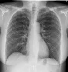
X-RAY CHEST PA VIEW
An X-ray chest PA view, also known as a posterior anterior chest X-rayAbout X-RAY CHEST PA VIEW
An X-ray chest PA view, also known as a posterior anterior chest X-ray, is a standard and routine imaging procedure used to examine the organs and structures within your chest cavity. Here's a comprehensive explanation of this X-ray technique:
Purpose of a Chest PA View X-ray:
A chest PA X-ray is a versatile tool used for a variety of diagnostic purposes, including:
- Diagnosing lung conditions: It can help identify abnormalities like pneumonia, infections, masses or nodules in the lungs, fluid collection around the lungs (pleural effusion), emphysema, and fibrosis.
- Evaluating chest injuries: Following a chest injury, an X-ray can reveal broken ribs, collapsed lung (pneumothorax), or injuries to the trachea (windpipe).
- Assessing heart health: While not as detailed as some other imaging techniques, a chest X-ray can provide a general view of the heart size and shape, and may indicate signs of heart failure.
- Screening purposes: In some cases, it might be used for screening individuals at risk for certain lung diseases, such as smokers.
Why PA view?
The PA view, where PA stands for posterior (back) anterior (front), means the X-ray beam originates from behind you and travels through your body to a detector placed in front of your chest. This positioning offers several advantages over an anterior-posterior (AP) view, where the X-ray beam comes from the front:
- Clearer view of the lungs: The PA view offers a more unobstructed view of the lungs, as the bulky structures like the breastbone and shoulder bones are spread apart during the procedure, allowing for better visualization behind them.
- Reduced heart size magnification: Since the X-ray beam travels through a lesser amount of tissue to reach the heart in a PA view, the heart size appears less magnified on the image compared to an AP view. This provides a more accurate assessment of actual heart size.
Procedure:
- Preparation: In most cases, no special preparation is required for a chest PA X-ray. You may be asked to remove any jewelry or clothing with metal fasteners that could interfere with the X-ray image. If you are pregnant, inform your doctor beforehand.
- Process: During the X-ray, you will likely stand in a booth or room with an X-ray machine. The technician will position you facing the detector plate and may ask you to raise your arms overhead or hold a breath for a moment while the X-ray image is captured. The entire procedure typically takes just a few minutes.
Benefits of a Chest PA View X-ray:
- Simple and quick: A chest PA X-ray is a simple, non-invasive, and outpatient procedure.
- Painless: The X-ray itself is painless, although you might feel some slight discomfort from positioning.
- Safe: The radiation dose from a chest X-ray is generally low.
- Effective for various conditions: It's a valuable tool for initial evaluation of a wide range of chest conditions.
Things to Consider:
- Radiation exposure: While the radiation dose is low, it's still important to be aware of it, especially for pregnant women or children undergoing multiple scans. Discuss the risks and benefits with your doctor.
- Limitations: A chest X-ray may not always provide a definitive diagnosis for all chest conditions. Sometimes, additional tests like a CT scan or ultrasound might be needed for a more detailed evaluation.
Overall, a chest PA view X-ray is a safe and effective initial imaging test for the chest cavity. If your doctor recommends a chest X-ray, discuss any concerns you may have about radiation exposure and ask about the reasons behind the recommendation, and whether a PA view is the most appropriate option.