CT THORAX
chest CT scanAbout CT THORAX
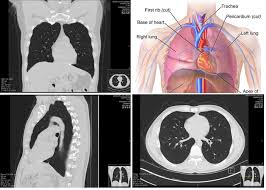
A CT thorax, also known as a chest CT scan, is a non-invasive imaging technique that uses X-rays to create detailed cross-sectional pictures of your chest cavity. This scan provides a comprehensive view of your internal organs in the chest, including:
- Lungs
- Heart
- Blood vessels
- Airways (trachea and bronchi)
- Esophagus (food pipe)
- Lymph nodes
- Muscles and bones of the chest wall
Why is a CT Thorax Performed?
Doctors utilize CT thorax scans for various diagnostic purposes, some of these include:
- Diagnosing lung conditions: A CT thorax can be highly effective in detecting lung abnormalities like pneumonia, blood clots in the lungs (pulmonary embolism), masses or nodules, infections, and chronic obstructive pulmonary disease (COPD).
- Evaluating chest injuries: Following a chest injury, a CT scan can reveal internal bleeding, fractures of the ribs or sternum, and damage to the lungs or other organs.
- Assessing heart health: A CT thorax can help visualize the heart, including its size, shape, and presence of abnormalities in the heart wall or valves. It can also detect blockages in the coronary arteries.
- Identifying tumors or masses: A CT thorax can help locate tumors or masses in the chest cavity, including lung cancer, esophageal cancer, or lymphoma.
- Staging cancer: If cancer is already diagnosed, a CT thorax can help determine the stage or extent of the cancer's spread.
- Planning treatment: The detailed images from a CT thorax can aid doctors in planning treatment approaches like surgery or radiation therapy.
What to Expect During a CT Thorax Scan:
- Preparation: In most cases, there's no special preparation required. You might be asked to remove any metal objects or clothing with metallic fasteners that could interfere with the scan images. If you are claustrophobic, inform your doctor beforehand. Depending on the scan type, you may be asked to drink a contrast solution beforehand to improve the visibility of certain structures.
- Procedure: During the scan, you'll lie down on a movable examination table that slides into the CT scanner, a large donut-shaped machine. Straps or headrests may be used to ensure you remain still during the scan. The CT scanner will rotate around your chest, taking multiple X-ray images from different angles. The entire procedure typically takes around 15 minutes, with the actual scanning time being just a few seconds.
Benefits of a CT Thorax Scan:
- Detailed images: A CT thorax scan provides highly detailed cross-sectional images of your chest cavity, allowing for a comprehensive examination of internal structures.
- Fast and accurate: The scan is relatively quick and provides accurate information for diagnosing various chest conditions.
- Non-invasive: No needles or injections are involved in a CT thorax scan.
Things to Consider:
- Radiation exposure: While the radiation dose from a CT scan is generally low, it is important to be aware of it, especially for pregnant women or children undergoing multiple scans. Discuss the risks and benefits with your doctor.
- Alternatives: Depending on the reason for the scan, your doctor might recommend alternative imaging tests like an X-ray (faster but less detailed) or an MRI scan (no radiation but more expensive and time-consuming).
Overall, a CT thorax scan is a valuable tool for diagnosing and evaluating a wide range of chest conditions. If your doctor recommends a CT thorax scan, discuss any concerns you may have about radiation exposure and ask about the reasons behind the recommendation.